Primed for the kill: occupying Bcl-2 to target death in acute myeloid leukaemia

Andrew Wei1,2*, Tse-Chieh Teh1
1The Australian Centre for Blood Diseases, Division of Blood Cancers, Monash University, Melbourne, Australia
2Department of Clinical Haematology, The Alfred Hospital, Commercial Road, Melbourne 3004, Australia

Citation: Wei A and Teh TC. Primed for the kill: occupying Bcl-2 to target death in acute myeloid leukaemia. BioDiscovery 2012; 6: 1; DOI: 10.7750/BioDiscovery.2012.6.1

Copyright: © 2012 Wei et al. This is an open-access article distributed under the terms of the Creative Commons Attribution License, which permits unrestricted use, provided the original authors and source are credited.

Received: November 24, 2012; Accepted: December 5, 2012; Available online /Published: December 7, 2012

Keywords: Bcl-2, AML, BH3-mimetic, apoptosis, Bim

*Corresponding Author: Andrew Wei, e-mail: a.wei@alfred.org.au

Conflict of Interests: No potential conflict of interest was disclosed by any of the authors.

Chemotherapy has been the mainstay of therapy for acute myeloid leukaemia (AML) for over five decades. Despite doses of cytarabine and anthracyclines approaching the limits of haematopoietic tolerance, half of all patients eventually relapse [1]. The situation is far worse for elderly patients, who are largely palliated because of the unfavourable risk-benefit ratio associated with intensive chemotherapy in older populations. Therapies tailored to specific sub-groups of AML have been remarkably effective, such as all-trans retinoic acid and arsenic for acute promyelocytic leukaemia, which have emerged as frontline therapies for this disease [2]. Other attempts to “target” AML, such as with FLT3 inhibitors have been less successful, especially in the setting of chemotherapy failure [3]. Gaining insights into the molecular basis of treatment failure will be a key objective for improving therapeutic outcomes of this aggressive blood cancer. For certain targeted therapies, such as FLT3 inhibitors, clinical resistance has been linked to emergence of “on-target” drug resistant mutations, which may be an important hurdle to the successful development of this drug class [4, 5]. The biological basis for treatment failure with non-targeted therapies, such as chemotherapy, has been far more complex to decipher.

The increasing queue of cytotoxic drugs failing to improve clinical outcomes in patients with relapsed and refractory AML indicates the existence of a fundamental mechanism conferring tolerance to genotoxic drugs within leukaemic cells. An attractive hypothesis was that multidrug resistance could be explained by the identification of drug efflux pumps on AML cells. This led to a generation of largely unsuccessful randomised clinical studies combining cytotoxic regimens to inhibitors of multidrug transporters, such as P-glycoprotein [6, 7]. The capacity for cytotoxic drugs to trigger apoptosis, or programmed cell death, has been extensively examined in model systems. Genotoxic drugs function in part by activating p53, and defects affecting the p53 pathway have an established role in counteracting the actions of DNA damaging agents [8]. In AML, p53 mutations only occur in a minority of cases, in particular, patients with complex cytogenetic karyotype [9]. Thus, defective p53 may not broadly explain the molecular basis for chemotherapy failure in AML. Mitochondrial mediated apoptosis leading to the activation of effector caspases is controlled by the Bcl-2 family of proteins [12]. Pro-apoptotic Bax and Bak are essential for inducing mitochondrial apoptosis. Cells lacking both Bax and Bak are resistant to chemotherapy mediated death [13]. Bax and Bak are kept inactive by a group of pro-survival Bcl-2 family members, including Bcl-2, Bcl-x, Bcl-w, Mcl-1 and A1. Cell stress signals, including chemotherapy, activate Bcl-2 homology-3 domain, termed “BH3-only” proteins [14]. BH3-only proteins bind and neutralise the effect of pro-survival Bcl-2 members, leading to downstream activation of pro-apoptotic Bax and Bak. Overexpression of pro-survival Bcl-2 and related proteins may cause resistance of multiple cytotoxic drugs to apoptosis [15]. Simple quantification of Bcl-2 expression may not accurately predict chemoresistance, as this fails to take into account whether prevailing Bcl-2 molecules are actually free to sequester BH3-only proteins or are already “occupied” and therefore rendered inert by pre-bound BH3-only protein. Another important nuance is that BH3-only proteins vary in their capacity to bind Bcl-2 targets. Bim, for instance, neutralises all anti-apoptotic Bcl-2 proteins potently, whereas Bad binding is limited to Bcl-2, Bcl-x and Bcl-w and Noxa can only bind Mcl-1 and A1 [16]. Thus, predicting the responsiveness of chemotherapy to mitochondrial apoptosis in cancerous and normal cells requires a readout that accounts for these multiple factors.

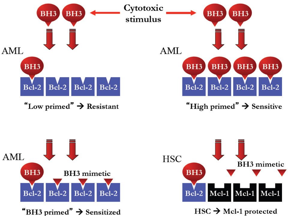
Thanh-Trang Vo and colleagues from Anthony Letai’s laboratory at Harvard have recently proposed that clinical responses to chemotherapy as well as long-term clinical outcomes may be determined functionally by assessing the capacity of BH3 peptides to induce mitochondrial depolarisation, a method they termed “BH3 profiling” [17]. An excess of BH3-only protein activity may lead to direct activation of Bax and Bak and ultimately, apoptosis [18]. The method assumes that the sensitivity of permeabilised cells to a quantum of exogenous BH3 peptide administered in vitro, is a direct reflection of the basal level of BH3 “priming” prevalent within the cell. Cells with greater levels of BH3-occupied Bcl-2 are therefore considered “highly primed” for death as measured by greater sensitivity to BH3 peptide. In contrast, an excess of BH3-unoccupied Bcl-2 proteins reflects a state of “low BH3 priming,” rendering the cell more resistant to mitochondrial depolarisation by BH3 peptide and consequently, the effects of some cytotoxic therapies (Figure 1). Compared to other attempts to gauge pro-survival activity in AML through expression analyses, BH3 profiling required no prior knowledge of the prevailing levels of pro-apoptotic or anti-apoptotic Bcl-2 family members. This may be especially useful in cases where Bcl-2 overexpression may lead to stabilisation and intracellular accumulation of Bim, resulting in relatively less unoccupied Bcl-2 than would be predicted by assessing Bcl-2 levels alone [19].

The Harvard team applied BH3 profiling to a large cohort of clinically annotated AML samples from the Dana-Farber and Memorial Sloan Kettering. From this work, they concluded that BH3 profiling was a determinant of initial response to induction chemotherapy, relapse after remission, and requirement for allogeneic bone marrow transplantation and that this information could be exploited for the personalization of therapy for AML. From a clinical perspective, how would this information be used? At presentation, most patients fit enough to receive chemotherapy will do so, because of the rapidly progressive nature of the disease. A large proportion of patients with low primed AML still achieve complete remission (CR), so that treatment would unlikely be deterred by this fact. Physicians must be as confident as possible that patients intended for stem cell transplantation are at high risk for relapse and that patients already in a cured state are not unintentionally transplanted. For patients achieving complete remission, BH3 profiling was able to distinguish a sub-group of approximately 20% of patients that were “cured” at that point and did not clinically relapse. All patients in this “cured” group had highly primed AML samples with a >60% response to 0.1μM BimBH3 peptide [17]. All patients with less responsive, or “low” primed AML were destined to relapse at some point. Thus, as a post-remission prognostic tool, the presence of highly BH3-primed AML may help to select a sub-group of patients that should not be recommended for allogeneic stem cell transplantation. To be broadly used however, a predictive test must be accurately validated and standardised in other laboratories. This remains a technically daunting task for the BH3 profiling assay, which requires the sample to be ficolled, permeabilised by digitonin, incubated with BH3 peptide and then stained with antibodies and mitochondrial dye to enable mitochondrial depolarization of blasts to be enumerated. Stable and reproducible controls for such a test would be a challenge to develop.

A more intriguing finding in the paper by Vo et al was the suggestion that the dominant pro-survival factor in human AML was Bcl-2, compared to Mcl-1 in normal hematopoietic stem cells (HSCs) [17]. Targeting Bcl-2 may therefore permit the selective destruction of AML cells, whilst sparing toxicity to normal HSCs, which would be protected by Mcl-1. Development of the BH3 small molecule mimetic ABT-737, which targets Bcl-2, Bcl-x and Bcl-w [20], and the clinical lead equivalent drug ABT-263 has been considered an unattractive prospect in AML, however, because of the critical role played by Bcl-x in platelet survival and the risk of exacerbating thrombocytopenia in a disease where bleeding is already a major cause of morbidity [21]. If the major pro-survival target in AML turns out to be Bcl-2, rather than Bcl-x, as suggested by Vo et al, this would allow small molecule BH3-only mimetics with selectivity against Bcl-2 (eg ABT-199), to target AML with a greater index of therapeutic safety than most cytotoxic drugs (Figure 1). One caveat is that Mcl-1 may be increased at the time of AML relapse, which may render AML resistant to Bcl-2 specific antagonists [22]. Emergence of Bcl-x overexpression may present another mechanism whereby resistant cancers may thwart the sustained success of Bcl-2 antagonists in the clinic [19].

Figure 1
Figure 1. Schematic illustration showing how low BH3 primed AML cells with an excess of un-occupied Bcl-2 may be more resistant to chemotherapy than a high BH3 primed cell. The level of BH3 priming may be enhanced by a BH3 mimetic targeting Bcl-2, making the cell more sensitive to chemotherapy. Haematopoietic stem cells (HSCs) expressing Mcl-1 are not targeted by BH3 mimetics with specificity for Bcl-2 and so toxicity to HSCs is not increased.

Previously, work by Glaser et al from the Walter and Eliza Hall Institute identified Mcl-1 as a critical survival factor in AML [23]. This group used inducible lentiviral vectors expressing BimBH3 variants to target survival in human AML cell lines and primary AML. In common with the Harvard group, a number of AML cell lines including MV4;11, HL-60, THP-1 and OCI-AML3 were examined, but with highly contradictory results. BH3 profiling revealed that these cell lines were more sensitive to Bad peptide (targets Bcl-2, Bcl-x and Bcl-w), compared to Noxa peptide (targets Mcl-1 and A1), which had little effect [17]. In contrast, Glaser et al found that all 4 cell lines were sensitive to expression of Bims2A (targets Mcl-1 only) and that MV4;11 and HL-60 cells were additionally sensitive to BimsBadBH3 (targets Bcl-2, Bcl-x and Bcl-w) [23, 24]. Experiments carried out by Glaser and colleagues using ABT-737 (a BadBH3 mimetic), were consistent with these findings [23]. This suggests that there may be important differences in the methodologies employed by these groups. BH3 profiling requires outer membrane permeabilisation, which may dilute the concentration of cytoplasmic proteins. Although Bcl-2 is membrane-localised, Bcl-x has both a cytosolic and membrane distribution [25, 26]. Whether the entire Bcl-x contribution to cell survival is adequately accounted for by the BH3-profiling technique is unclear. A panel of cell permeable small molecules selectively targeting each of Bcl-2, Bcl-x or Mcl-1 alone, would represent a perfect “toolkit” for probing cell survival dependence in primary human AML cells. To this end, a potent Bcl-2 selective inhibitor has already been developed (ABT 199) and prototype Mcl-1 selective inhibitors are emerging [27].

Next generation sequencing has shown that hundreds of coding mutations potentially exist within each AML genome [28]. A reductionist approach targeting the collective impact of these altered genomes on the core survival apparatus in AML, therefore, has great therapeutic appeal. The work by Vo et al suggests that targeting Bcl-2 may lie at the heart of this strategy. Fortunately, a lead clinical compound called ABT-199 (Genentech) has already been developed to lead this charge. The activity of BH3-mimetics in AML, such as ABT-737, may be enhanced through combination with FLT3, MEK or MDM2 inhibitors, cytotoxic drugs or demethylating agents [29]. Several of these drugs may augment levels of Bim protein, thereby assisting ABT-737 to overwhelm Bcl-2 and initiate apoptosis. Unlike ABT-737, which targets Bcl-x and causes thrombocytopenia, the action of ABT-199 is limited to Bcl-2, thus avoiding this unwanted risk. Pre-clinical studies to verify the activity of ABT-199 in AML and to examine patterns of potential resistance are urgently awaited. For the large community of AML sufferers in desperate need of more effective therapies, however, the activation of clinical trials to tackle Bcl-2 in AML cannot come quickly enough.

Acknowledgments

We wish to acknowledge funding support received from the Leukaemia Foundation of Australia, the Victorian Cancer Agency and the National Health and Medical Research Council of Australia.

References

  • Fernandez H, Sun Z, Yao X, Litzow M, Luger S, Paietta E, et al. Anthracycline dose intensification in acute myeloid leukemia. The New England Journal of Medicine 2009; 361: 1249-1259.
    Reference Link
  • Iland H, Bradstock K, Supple S, Catalano A, Collins M, Hertzberg M, et al. All-trans-retinoic acid, idarubicin, and IV arsenic trioxide as initial therapy in acute promyelocytic leukemia (APML4). Blood 2012; 120: 1570-1580.
    Reference Link
  • Levis M, Ravandi F, Wang E, Baer M, Perl A, Coutre S, et al. Results from a randomized trial of salvage chemotherapy followed by lestaurtinib for patients with FLT3 mutant AML in first relapse. Blood 2011; 117: 3294-3301.
    Reference Link
  • Smith C, Wang Q, Chin C-S, Salerno S, Damon L,1 Levis M, et al. Validation of ITD mutations in FLT3 as a therapeutic target in human acute myeloid leukaemia. Nature 2012; 485: 260-263.
  • Man C, Fung T, Ho C, Han H, Chow H, Ma A, et al. Sorafenib treatment of FLT3-ITD+ acute myeloid leukemia: favorable initial outcome and mechanisms of subsequent non-responsiveness associated with a D835 mutation. Blood 2012; 119: 5133-5143.
    Reference Link
  • Becton D, Dahl G, Ravindranath Y, Chang M, Behm F, Raimondi S, et al. Randomized use of cyclosporin A (CsA) to modulate P-glycoprotein in children with AML in remission: Pediatric Oncology Group Study 9421. Blood 2006; 107: 1315-1324.
    Reference Link
  • van der Holt B, Löwenberg B, Burnett A, Knauf W, Shepherd J, Piccaluga P, Ossenkoppele G, et al. The value of the MDR1 reversal agent PSC-833 in addition to daunorubicin and cytarabine in the treatment of elderly patients with previously untreated acute myeloid leukemia (AML), in relation to MDR1 status at diagnosis. Blood 2005; 106: 2646-2654.
  • Li R, Sutphin PD, Schwartz D, Matas D, Almog N, Wolkowicz R et al. Mutant p53 protein expression interferes with p53-independent apoptotic pathways. Oncogene 1998; 16: 3269-3277.
    Reference Link
  • Stirewalt DL, Kopecky KJ, Meshinchi S, Appelbaum FR, Slovak ML, Willman CL, et al. FLT3, RAS, and TP53 mutations in elderly patients with acute myeloid leukemia. Blood 2001; 97: 3589-3595.
    Reference Link
  • Fenaux P, Jonveaux P, Quiquandon I, Laï JL, Pignon JM, Loucheux-Lefebvre MH, et al. P53 gene mutations in acute myeloid leukemia with 17p monosomy. Blood 1991; 78: 1652-1657.
  • Haferlach C, Dicker F, Herholz H, Schnittger S, Kern W, Haferlach T. Mutations of the TP53 gene in acute myeloid leukemia are strongly associated with a complex aberrant karyotype. Leukemia 2008; 22: 1539-1541.
    Reference Link
  • Strasser A, Cory S, Adams JM. Deciphering the rules of programmed cell death to improve therapy of cancer and other diseases. The EMBO Journal 2011; 30: 3667-3683.
    Reference Link
  • Cheng E, Wei M, Weiler S, Flavell R, Mak T, Lindsten T, et al. BCL-2, BCL-XL Sequester BH3 Domain-Only Molecules Preventing BAX-and BAK-Mediated Mitochondrial Apoptosis. Molecular Cell 2001; 8: 705-711.
    Reference Link
  • Villunger A, Michalak EM, Coultas L, Müllauer F, Böck G, Ausserlechner M, et al. p53-and drug-induced apoptotic responses mediated by BH3-only proteins puma and noxa. Science Signalling 2003; 302: 1036.
  • Schmitt CA, Lowe SW. Bcl-2 Mediates Chemoresistance in Matched Pairs of Primary Eμ-myc Lymphomas in Vivo. Blood Cells, Molecules, and Diseases 2001; 27: 206-216.
    Reference Link
  • Chen L, Willis SN, Wei A, Smith BJ, Fletcher JI, Hinds MG, et al. Differential targeting of prosurvival Bcl-2 proteins by their BH3-only ligands allows complementary apoptotic function. Mol Cell 2005; 17: 393-403.
    Reference Link
  • Vo, T, Ryan J, Carrasco R, Neuberg D, Rossi D, Stone RM, et al. Relative Mitochondrial Priming of Myeloblasts and Normal HSCs Determines Chemotherapeutic Success in AML. Cell 2012; 151: 344-355.
    Reference Link
  • Wei MC, Zong WX, Cheng EH, Lindsten T, Panoutsakopoulou V, Ross AJ, et al. Proapoptotic BAX and BAK: a requisite gateway to mitochondrial dysfunction and death. Science Signalling 2001; 292: 727-730.
  • Mérino D, Khaw SL, Glaser SP, Anderson DJ, Belmont LD, Wong C, et al. Bcl-2, Bcl-xL, and Bcl-w are not equivalent targets of ABT-737 and navitoclax (ABT-263) in lymphoid and leukemic cells. Blood 2012; 119: 5807-5816.
  • Oltersdorf T, Steven W. Elmore SW, Shoemaker AR, Armstrong RC, Augeri DJ, Belli BA, et al. An inhibitor of Bcl-2 family proteins induces regression of solid tumours. Nature 2005; 435: 677-681.
  • Mason K, Vandenberga CJ, Scotta CL, Wei AH, Corya S, Huanga D, et al. In vivo efficacy of the Bcl-2 antagonist ABT-737 against aggressive Myc-driven lymphomas. Proceedings of the National Academy of Sciences 2008; 105: 17961-17966.
    Reference Link
  • Reed, J. C. Bcl-2 family proteins: regulators of apoptosis and chemoresistance in hematologic malignancies. Semin Hematol 1997; 34: 9-19.
  • Glaser SP, Lee EF, Trounson E, Bouillet P, Wei A, Fairlie D, et al. Anti-apoptotic Mcl-1 is essential for the development and sustained growth of acute myeloid leukemia. Genes & Development 2012; 26: 120-125.
    Reference Link
  • Lee EF, Czabotar P, van Delft MF, Michalak EM, Boyle MJ, Willis SN, et al. A novel BH3 ligand that selectively targets Mcl-1 reveals that apoptosis can proceed without Mcl-1 degradation. The Journal of Cell Biology 2008; 180: 341-355.
    Reference Link
  • Hockenbery D, Nuñez G, Milliman C, Schreiber RD, Korsmeyer SJ. Bcl-2 is an inner mitochondrial membrane protein that blocks programmed cell death. Nature 1990; 348: 334-336.
    Reference Link
  • Hsu YT, Wolter KG, Youle RJ. Cytosol-to-membrane redistribution of Bax and Bcl-XL during apoptosis. Proceedings of the National Academy of Sciences 1997; 94: 3668-3672.
    Reference Link
  • Doi K, Li R, Sung SS, Wu H, Liu Y, Manieri W, et al. Discovery of marinopyrrole A (maritoclax) as a selective Mcl-1 antagonist that overcomes ABT-737 resistance by binding to and targeting Mcl-1 for proteasomal degradation. Journal of Biological Chemistry 2012; 287: 10224-10235.
    Reference Link
  • Ding L, Ley TJ, Larson DE, Miller CA, Koboldt DC, Welch JS, et al. Clonal evolution in relapsed acute myeloid leukaemia revealed by whole-genome sequencing. Nature 2012; 481: 506-510.
    Reference Link
  • Kohl TM, Hellinger C, Ahmed F, Buske C, Hiddemann W, Bohlander SK, et al. BH3 mimetic ABT-737 neutralizes resistance to FLT3 inhibitor treatment mediated by FLT3-independent expression of BCL2 in primary AML blasts. Leukemia 2007; 21: 1763-1772.
    Reference Link
  • Kojima K, Konopleva M, Samudio IJ, Schober WD, Bornmann WG, Andreeff M, et al. Concomitant inhibition of MDM2 and Bcl-2 protein function synergistically induce mitochondrial apoptosis in AML. Cell Cycle 2006; 5: 2778-2786.
    Reference Link
  • Tsao T, Shi Y, Kornblau S, Lu H, Konoplev S, Antony A, et al. Concomitant inhibition of DNA methyltransferase and BCL-2 protein function synergistically induce mitochondrial apoptosis in acute myelogenous leukemia cells. Annals of Hematology 2012; 91: 1-10.
    Reference Link
  • Konopleva M, Milella M, Ruvolo P, Watts JC, Ricciardi MR, Korchin B, et al. MEK inhibition enhances ABT-737-induced leukemia cell apoptosis via prevention of ERK-activated MCL-1 induction and modulation of MCL-1/BIM complex. Leukemia 2011; 26: 778-787.
    Reference Link