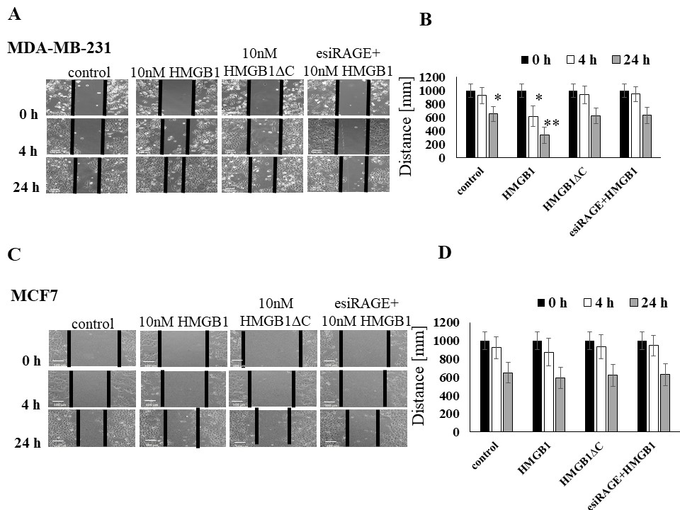
The effect of HMGB1 and HMGB1∆C recombinant proteins on the mobility of control and cancer cells with RAGE-expressed and RAGE silenced protein, measured by wound healing assay. Panel A. 90% confluent monolayers of MDA-MB-231 cells were scratch wounded. Images of wounded monolayer were taken at times 0 h, 4 h and 24 h. Cells are incubated with recHMGB1 and recHMGB1∆C proteins (10 nM) with RAGE expressing cells and with silencing RAGE cells. The vertical lines indicate the wound edge. Panel B and D. Graphical representation of cell movement by measuring the distance between wound healing lines. The values represent the means ± SD (n=4). Statistical significance was calculated by Student’s t test, and * P-values ≤0.05 were considered to indicate statistically significant results, **p-values ≤0.001. Bars indicate 20 μm. Panel C. 80% confluent monolayers of MCF 7 cells were scratch wounded. Images of wounded monolayer were taken at times 0 h, 4 h and 24 h. Cells are incubated with recHMGB1 and recHMGB1∆C proteins (10 nM) with RAGE expressing cells and with silencing RAGE cells. The vertical lines indicate the wound edge and plotted the distance between wound healing lines.