Up-regulation of Gr1 + CD11b + cell population in the spleen of NaClO-administered mice works to repair skin wounds

Mayu Hara1,2, Naomi Nishio1, Sachiko Ito1, Masashi Akiyama2 and Ken-ichi Isobe1*
1 Department of Immunology, Nagoya University Graduate School of Medicine, Nagoya, Aichi, 466-8550, Japan
2 Department of Dermatology, Nagoya University Graduate School of Medicine, Nagoya, Aichi, 466-8550, Japan

Abstract

In wound healing, early infiltration of neutrophils followed by macrophage infiltration are important defense mechanisms for repair of tissue damage. Here we examined the effects of neutrophils on wound healing. Administration of sodium hypochlorite (NaClO) to mouse skin induces neutrophil recruitment to the wound site and repeated administration of NaClO was shown to prolong wound healing. Examination of the spleens of mice whose wounds were repeatedly treated with NaClO, showed that GR-1+CD11b+ cells were up regulated in the recovery phase of wounding. Many of the GR-1+CD11b + cells in the mouse bone marrow were neutrophils, as indicated by a ring-shaped nucleus, and some of the cells were immature myeloid-lineage cells. GR-1+CD11b+ cells from bone marrow were sorted and injected intravenously to syngeneic Imprinting Control Region (ICR) mice. The mice that received GR-1+CD11b+ cells recovered faster than the mice injected with the control, phosphate buffer saline (PBS).

Citation: Hara M, Nishio N, Ito S, Akiyama M, Isobe K. Up-regulation of Gr1+CD11b+ cell population in the spleen of NaClO- administered mice works to repair skin wounds Biodiscovery 2012; 2: 1; DOI: 10.7750/BioDiscovery.2012.2.1

Copyright: © 2012 Hara et al. This is an open-access article distributed under the terms of the Creative Commons Attribution License, which permits unrestricted use, provided the original authors and source are credited.

Received: June 22, 2012; Accepted: August 1, 2012; Available online /Published: August 6, 2012

Keywords: neutrophils, wound healing, GR-1+CD11b+ cells

Abbreviations: checkpoint: mitotic spindle assembly checkpoint

*Corresponding Author: Isobe K.; email: kisobe@med.nagoya-u.ac.jp

Conflict of Interests: The authors declare no competing financial interests.

Introduction

Neutrophils and macrophages engulf not only microorganisms but also damaged tissue and degrade these materials in the phagolysosome [1]. In wound healing, early infiltration of neutrophils followed by macrophage infiltration are important defense mechanisms for repair of tissue damage. Infiltrating neutrophils clear the wounded area of foreign particles and bacteria and are then extruded with the eschar or phagocytosed by macrophages. Subsequent to neutrophil invasion, monocytes infiltrate the wound site and become activated macrophages that release growth factors such as platelet-derived growth factor and vascular endothelial growth factor, which initiate the formation of granulation tissue [6,7]. Recently, it has been shown that damage associated molecular patterns (DAMPs) from injured tissues cause innate cell activation [8]. Wounding induces death of tissue cells, which occurs upon the loss of plasma membrane integrity, thereby allowing the escape of intracellular material from the cells including heat shock proteins (HSPs), high-mobility group box protein 1 (HMGB1), ATP and uric acid. These intracellular materials, along with extracellular materials such as hyaluronan and heparin sulphate produced by proteolytic activity of dying cells, bind to pattern recognition receptors (PRRs) of resident macrophages and dendritic cells. Reactive oxygen species (ROS) are also produced upon wounding and have been shown to activate the inflammasome [8]. Activated cells produce inflammatory cytokines and chemokines, especially IL-8, which induces the migration of neutrophils to wounds. Migrated neutrophils are also activated by DAMPs and produce inflammatory cytokines and several toxic enzymes, which have positive and negative effects on would healing.

The function of macrophages in wound healing has been well studied. Classically activated macrophages (M1 macrophages) mediate defense of the host from a variety of bacteria, protozoa and viruses, and have roles in antitumor immunity. Alternatively activated macrophages (M2 macrophages) have anti-inflammatory functions and regulate wound healing. 'Regulatory' macrophages can secrete large amounts of interleukin-10 (IL-10) in response to Fc receptor-γ ligation [11, 12]. In contrast to pro-inflammatory and anti-microbial M1 macrophage responses, M2 macrophages exhibit potent anti-inflammatory activity and have important roles in wound healing and fibrosis [13, 14]. However, there are contradictory reports about the functions of neutrophils in wound healing. Classically, depletion of neutrophils by antisera has no effects on wound healing, although the depletion of macrophages results in a failure of debridement [18, 16]. We reported previously that the depletion of neutrophils by anti-Gr-1 antibody dramatically delayed wound healing in aged mice, although the depletion of neutrophils in young mice had less effect on the kinetics of wound healing [17].

We have recently shown that Gr1+CD11b+ cells increased in the spleen of dextran sulphate sodium (DSS)-treated mice during the recovery phase and splenectomy worsened the colitis [18]. DSS-derived splenic Gr1+CD11b+cells were administered intravenously to recipient (C57BL/6) mice during the early phase of DSS treatment. The transplanted splenic DSS-induced Gr1+CD11b+cells improved DSS-induced colitis and promoted efficient colonic mucosal healing [19]. As many of the Gr1+CD11b+ cells are neutrophils, they may play a role in wound repair.

During infection, the innate immune system functions to kill microorganisms that induce tissue damage. Once immune cells clear the microorganisms, tissue regeneration occurs and damaged tissues are repaired. However, if the immune system fails to clear all the microorganisms such as hepatitis C virus, repeated stimulation by the virus will induce tissue destruction and finally organ failure. We hypothesize that although immune cells repair single damage, continuous stimulation of the immune system may induce chronic inflammation and induce tissue degeneration. Here we examined the function of neutrophils in wound healing by single or repeated stimulation of oxidative tissue damage by ROS-producing materials.

Results

A single application of NaClO induces migration of neutrophils to a wound site

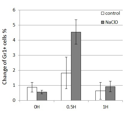
As it has been shown that ROS induces neutrophil migration to wound sites in zebrafish [20], we asked whether NaClO induces migration of neutrophils to wound sites in mice. A single drop of NaClO was administered to a wound and migration of neutrophils to the wound site was examined. The single drop of NaClO to the wound opening induced higher neutrophil migration as compared to a simple wound. Neutrophils migrated to the wound within 0.5 hr and neutrophil number decreased 1h after wounding (Fig. 1).

Figure 1
Figure 1. Greater migration of neutrophils upon a single application of NaClO.The dorsal skins of 2 month-old female ICR mice (n=3) were wounded by puncturing through two layers of skin with a sterile disposable 3mm biopsy punch. NaClO (available Cl 2.5%) was applied once to the wound site. The wounds and their surrounding areas (including the scab and epithelial margins) were removed for FACS analysis using a sterile disposable biopsy punch with a diameter of 6 mm at the indicated time points. Single cell suspensions were obtained by passing through stainless mesh and washed with PBS. Biopsied tissues were squeezed by rubber in PBS, passed through stainless mesh, and washed with PBS. The cell suspensions were stained with FITC-labeled-antiGR-1 and analyzed by FACS sorting.

Repeated application of NaClO prolongs wound healing

We thought that repeated exposure of ROS might affect wound healing. If wounds are repeatedly treated with NaClO, neutrophils must be continuously provided by an organ from which they originate. Spleen and bone marrow in mice have hematopoietic stem cells, which provide new neutrophils. GR-1+CD11b+ cells are a mixed population, containing precursors of neutrophils in addition to mature neutrophils. As shown in Fig. 2, application of NaClO for 5 days strongly damaged the skin tissue of mice and delayed wound healing. Upon examination of the spleens of mice that were subject to repeated application of NaClO, GR-1 +CD11b+ positive cells were shown to be up-regulated during the recovery phase of wounding (Fig. 3).
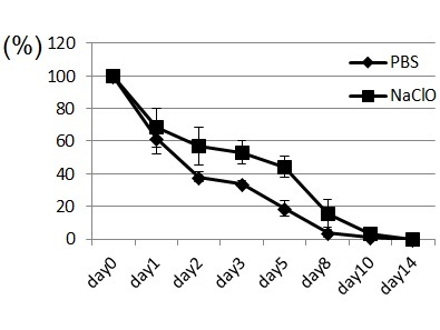
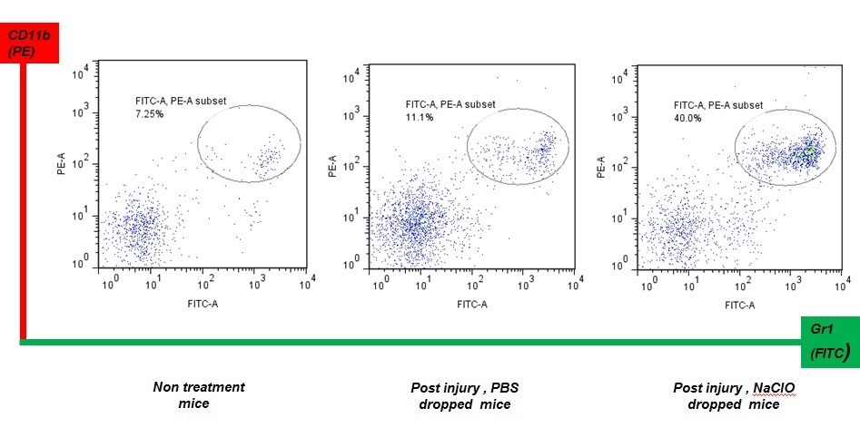
Figure 2
Figure 2. Wound repair is delayed by the continuous application of NaClO. The dorsal skin of 14 week-old female ICR mice (n=5) were punctured through two layers of skin with a sterile disposable 3mm biopsy punch. NaClO (available Cl 1%) was applied once a day to the wounded site for 5 days.Changes in the area were calculated as a percentage of the initial wound area. Changes in percentages of wound areas at each time point were compared to the initial wound area. Data shown are the mean ratio ± SE of four separate experiments
Figure 3
Figure 3. Up-regulation of GR-1+ CD11b+ cells in the spleen of NaClO-treated mice. The dorsal skins of 2 month-old (at sacrifice) female ICR mice were punctured through two layers of skin with a sterile disposable 3mm biopsy punch. NaClO (available Cl 2.5%) was applied once a day to the wound site for 5 days. The spleen of NaClO- administered mice was removed 11 days after the last day of NaClO application. The cell suspensions were stained with FITC-labeled-antiGR-1 and PE-labeled anti-CD11b and analyzed by FACS sorting.

Transfer of GR-1+CD11b+ positive cells induces early recovery from wounding

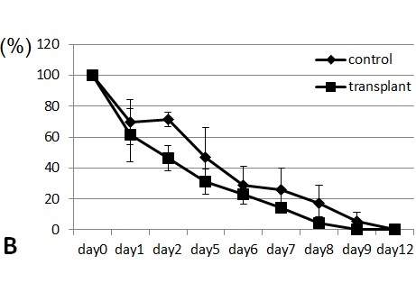
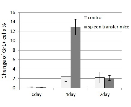
Spleen cells from wounded mice that had received 11-day treatments with NaClO were removed and transplanted intravenously to syngeneic ICR mice. The transplanted mice were subsequently wounded and recovery from the wounds was monitored. Mice that received the transplanted spleen cells from NaClO-administered mice recovered faster than mice that received the control spleen ( Figure 4). GR-1+CD11b+ -positive cells from the bone marrow of untreated ICR mice were FACS sorted, cytospinned, and stained by Gimsa. Many of the GR-1+CD11b+ cells were shown to be neutrophils, as indicated by their ring-shaped nucleus. Some of the cells were immature myeloid-lineage cells (Fig. 5A). The sorted GR-1+CD11b+ cells were injected into syngeneic ICR mice intravenously. Mice that received the transplanted GR-1+CD11b + cells recovered faster than the mice that received the control PBS. (Fig.5B). Then we examined the appearance of neutrophils at the wounded sites. We found that GR-1+ neutrophils appeared earlier and higher in the wounded tissues of the mice that received the spleen cells from NaClO - administered mice, than in the control mice (Fig.6).
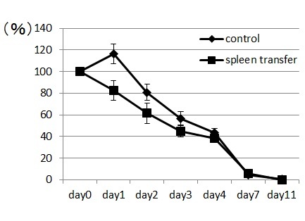
Figure 4
Figure 4. Transfer of the spleen of NaClO (available Cl 2.5%)-treated mice induces early wound recovery.The dorsal skin of an 8 week-old (at sacrifice) female ICR mouse (n=1) was punctured through two layers of skin with a sterile disposable 3mm biopsy punch. NaClO was applied once a day to the wound site for 5 days. The spleen of NaClO-administered mice was removed at 11 days after the last day of NaClO application. The cell suspensions (1x10⁷ cells /mouse) were transferred to ICR mice (8 weeks old, female, n=4) just before punch biopsy. Shown are changes in the percentage of wound areas at each time point in comparison to the initial wound area. Data shown are the mean ratio ± SE of four separate experiments.
Figure 5a Figure 5b
Figure 5. Transfer of GR-1+CD11b+ cells in bone marrow cells induces early wound recovery. Bone marrow cells were collected from a mouse (ICR, female, 18weeks old, n=1). The cell suspensions were stained with FITC-labeled-anti-GR-1 and PE-labeled anti-CD11b, and GR-1+CD11b+ cells were FACS sorted. A. Cytospin and Gimsa staining of sorted cells. B. Sorted cells (1.3x10⁶ cells/mouse) were injected into ICR mice (female, 10 weeks old, n=3) just before wounding. Shown are changes in the percentage of wound areas at each time point in comparison to the initial wound area. Data shown are the mean ratio ± SE of four separate experiments.
Figure 3
Figure 6. Early appearance and higher numbers of neutrophils in transferred mice. The dorsal skins of 2 month-old (at sacrifice) female ICR mouse were punctured through two layers of skin with a sterile disposable 3mm biopsy punch. NaClO (available Cl 2.5%) was applied once a day to the wound site for 5 days. The spleen of NaClO-administered mice (ICR, female, 8weeks old, n=3) was removed 11 days after the last day of NaClO application. The cell suspensions (1x10 7 cells /mouse) were transferred to ICR mice (8 weeks old, female, n=4) just before punch biopsy. Tissues were collected from the wound site by punch biopsy (diameter 6mm) at days 0, 1, and 2. Single cell suspensions were obtained by passing through stainless mesh and washed with PBS. Biopsied tissues were mechanically disrupted with rubber in PBS, passed through stainless mesh, and washed with PBS. The cell suspensions were stained with FITC-labeled antiGR-1 and analyzed by FACS sorting.

Discussion

We have shown here that application of NaClO to wounded tissue induces neutrophil migration to the wound site. This migration was found to be transient after a single application of NaClO. Thus, we applied NaClO for 5 days to wound sites to induce continuous neutrophil migration. Neutrophils produce ROS, lytic enzymes and antimicrobial peptides, which facilitates the killing of microorganisms and clear damaged tissues [21, 22]. These products of neutrophils also produce cytokines, which induce local inflammation, manifested by swelling and fever. Once the noxious stimuli are withdrawn, local inflammation is down- regulated and tissue recovery can occur. However, repeated exposure to tissue-damaging stimuli may induce chronic neutrophil migration and thus, chronic inflammation. When we examined the spleens of mice that had received continuous application of NaClO, the population of GR-1+CD11b+ cells was up-regulated. The population of GR-1+CD11b+ cells contains mainly neutrophils but also some myeloid-precursor cells as shown by cytospin (Fig. 5A). We conclude that these populations are a mixture of myeloid-precursor cells and differentiated neutrophils. Repeated exposure of the wound to ROS may induce neutrophil-precursors, together with mature neutrophils, in the spleen in order to supply sufficient numbers of neutrophils to the damaged tissue. These results are correlated with our previous work, which showed up-regulation of the GR-1+CD11b+ population in the spleen of DSS-treated mice. DSS also produces ROS in the intestine and induces neutrophil migration [18,19]. The transplantation of GR-1+CD11b+ cells in the early phase of wound healing induces early recovery of wound healing. We detected higher amounts of neutrophils at wound sites upon transplantation of GR-1+CD11b+ cells. These results indicate that the transient increase of neutrophils at damaged sites facilitates wound healing and may function in the clearing of damaged wound tissues.

Servettaz et al. developed the murine model of dermal sclerosis by repeated injection of ROS-inducing agents, including NaClO [23]. We have also reported that the ROS-producing agent Bleomycin induces systemic sclerosis [24], although Servettaz et al. and our group have not examined the contribution of neutrophils to disease progression. ROS-producing agents are known to induce local neutrophil recruitment and repeated administration of ROS may induce persistent neutrophil recruitment to the application site. Although further molecular and cellular analyses of this process are needed, the results in this paper seem to indicate a role for neutrophils in host defense wound healing. In the future, incurable wounds may be treated by injection of the patient's own neutrophils that have been differentiated from the patient’s own stem cells (iPSCs or myeloid-lineage stem cells) in vitro.

Materials and Methods

Mice

8-18 week-old ICR mice were originally purchased from SLC Japan and maintained at the Animal Research Facility at Nagoya University Graduate School of Medicine under specific pathogen-free conditions and used according to institutional guidelines.

Excision wound preparation and macroscopic examination

Avertin (0.2ml per gram of body weight) was used as anesthesia during the wound generation and skin removal. After shaving and extensive cleaning with 70% ethanol, the dorsal skin was picked up at the midline and holes were punched through two layers of skin with a sterile disposable biopsy punch (diameter 3 mm; Kai Industries, Tokyo, Japan). At one side NaClO (available Cl 1-2.5%) was dropped and on the other side PBS was dropped as a control. This procedure generated two excisional full-thickness wounds on each side of the midline. The same procedure was repeated 3-5 times, generating 6-10 wounds on each animal. Each wound site was digitally photographed at the indicated time intervals, and wound areas were delineated using Excel software. Changes in wound areas were expressed as the proportion of the change in wound area relative to the initial wound area. In some experiments, wounds and their surrounding areas (including the scab and epithelial margins) were removed for further analysis using a sterile disposable biopsy punch with a diameter of 6 mm (Kai Industries) at the indicated time points.

FACS analysis and cell sorting

Wound tissue and spleen were gently clashed by rubber. Single cell suspensions were obtained by passing through stainless mesh and washed with PBS. Cells were treated with FITC-labeled anti-mouse Gr-1 or PE-labeled anti-mouse CD11b (BD Pharmingen). Cells were sorted with a FACS Aria (BD Biosciences) and data analyzed with FlowJo software (Tree Star).

Acknowledgements

We thank M. Tanaka for technical assistance with flow cytometry and N. Oiwa for secretarial work. This work was supported by the Ministry of Education, Science, Technology, Sports and Culture, Japan and the Global Centers of Excellence Program of Nagoya University.

References

  • Gordon S, Taylor PR. Monocyte and macrophage heterogeneity. Nat Rev Immunol 2005; 5(12): 953-64.
    Reference Link
  • Geissmann F, Gordon S, Hume DA, Mowat AM, Randolph GJ. Unravelling mononuclear phagocyte heterogeneity. Nat Rev Immunol 2010; 10(6): 453-60.
    Reference Link
  • Ricardo SD, van Goor H, Eddy AA. Macrophage diversity in renal injury and repair. J Clin Invest 2008; 118(11): 3522-30.
    Reference Link
  • Wynn TA, Barron L. Macrophages: master regulators of inflammation and fibrosis. Semin Liver Dis 2010; 30(3): 245-57.
    Reference Link
  • Murray PJ, Wynn TA. Protective and pathogenic functions of macrophage subsets. Nat Rev Immunol 2011; 11(11): 723-37.
    Reference Link
  • Singer AJ, Clark RA. Cutaneous wound healing. N Engl J Med 1999; 341(10): 738-46.
    Reference Link
  • Martin P, Leibovich SJ. Inflammatory cells during wound repair: the good, the bad and the ugly. Trends Cell Biol 2005; 15(11): 599-607.
    Reference Link
  • Chen GY, Nu?ez G. Sterile inflammation: sensing and reacting to damage. Nat Rev Immunol 2010; 10(12): 826-37.
    Reference Link
  • Newton K, Dixit VM. Signaling in innate immunity and inflammation. Cold Spring Harb Perspect Biol 2012; 4(3).
    Reference Link
  • Rock KL, Latz E, Ontiveros F, Kono H. The sterile inflammatory response. Annu Rev Immunol 2010; 28: 321-42.
    Reference Link
  • Sutterwala FS, Noel GJ, Clynes R, Mosser DM. Selective suppression of interleukin-12 induction after macrophage receptor ligation. J Exp Med 1997; 185(11): 1977-85.
    Reference Link
  • Sutterwala FS, Noel GJ, Salgame P, Mosser DM. Reversal of proinflammatory responses by ligating the macrophage Fcgamma receptor type I. J Exp Med 1998; 188(1): 217-22.
    Reference Link
  • Wynn TA. Fibrotic disease and the T(H)1/T(H)2 paradigm. Nat Rev Immunol 2004; 4(8): 583-94.
    Reference Link
  • Xiao W, Hong H, Kawakami Y, Lowell CA, Kawakami T. Regulation of myeloproliferation and M2 macrophage programming in mice by Lyn/Hck, SHIP, and Stat5. J Clin Invest 2008; 118(3): 924-34.
  • Simpson DM, Ross R. The neutrophilic leukocyte in wound repair a study with antineutrophil serum. J Clin Invest 1972; 51(8): 2009-23.
    Reference Link
  • Leibovich SJ, Ross R. The role of the macrophage in wound repair. A study with hydrocortisone and antimacrophage serum. Am J Pathol 1975; 78(1): 71-100.
  • Nishio N, Okawa Y, Sakurai H, Isobe K. Neutrophil depletion delays wound repair in aged mice. Age (Dordr) 2008; 30(1): 11-9.
    Reference Link
  • Zhang R, Ito S, Nishio N, Cheng Z, Suzuki H, Isobe K. Up-regulation of Gr1+CD11b+ population in spleen of dextran sulfate sodium administered mice works to repair colitis. Inflamm Allergy Drug Targets 2011; 10(1): 39-46.
    Reference Link
  • Zhang R, Ito S, Nishio N, Cheng Z, Suzuki H, Isobe KI. Dextran sulphate sodium increases splenic Gr1(+)CD11b(+) cells which accelerate recovery from colitis following intravenous transplantation. Clin Exp Immunol 2011; 164(3): 417-27.
    Reference Link
  • Niethammer P, Grabher C, Look AT, Mitchison TJ. A tissue-scale gradient of hydrogen peroxide mediates rapid wound detection in zebrafish. Nature 2009; 459(7249): 996-9.
    Reference Link
  • Nathan C. Neutrophils and immunity: challenges and opportunities. Nat Rev Immunol 2006; 6(3): 173-82.
    Reference Link
  • Borregaard N. Neutrophils, from marrow to microbes. Immunity 2010; 33(5): 657-70.
    Reference Link
  • Servettaz A, Goulvestre C, Kavian N, Nicco C, Guilpain P, Ch?reau C et al. Selective oxidation of DNA topoisomerase 1 induces systemic sclerosis in the mouse. J Immunol 2009; 182(9): 5855-64.
  • Ishikawa H, Takeda K, Okamoto A, Matsuo S, Isobe K. Induction of autoimmunity in a bleomycin-induced murine model of experimental systemic sclerosis: an important role for CD4+ T cells. J Invest Dermatol 2009; 129(7): 1688-95.
    Reference Link