To see an interphase chromosome or: How a disease can be associated with specific nuclear genome organization

Ivan Y. Iourov
1 Mental Health Research Center, Russian Academy of Medical Sciences, 119152 Moscow, Russia
2 Institute of Pediatrics and Children Surgery, Ministry of Health of the Russian Federation, 125412 Moscow, Russia

Abstract

The last hierarchical level of cellular genome organization is the spatial arrangement of chromosomes within the nuclear space. Despite of high regulatory potential and functional implications, issues concerning nuclear organization at chromosomal level are rarely addressed because of limitations in visualizing interphase chromosomes. The problem is especially seen when an attempt to associate specific patterns of nuclear genome organization with a pathological condition is made. Fortunately, advances in molecular cytogenetics have provided for a solution to visualize chromosomes in interphase nuclei at molecular resolution. A study in this issue of BioDiscovery shows the way of how to identify interphase chromosome architecture at molecular resolutions and demonstrates the involvement of specific nuclear genome organization in generating a cancer-causing chromosomal aberration (translocation between chromosomes 8 and 21 in acute myelogenous leukemia). Authors’ findings suggest interphase molecular cytogenetic techniques (i.e. interphase chromosome-specific multicolor banding or ICS-MCB) to be required to perform studies regarding nuclear genome organization at chromosomal level and its role in disease pathogenesis.

Citation: Iourov I. To see an interphase chromosome or: How a disease can be associated with specific nuclear genome organization. BioDiscovery 2012; 4: 5; DOI: 10.7750/BioDiscovery.2012.4.5

Copyright: © 2012 Iourov. This is an open-access article distributed under the terms of the Creative Commons Attribution License, which permits unrestricted use, provided the original authors and source are credited.

Received: October 29, 2012; Accepted: October 30, 2012; Available online /Published: October 31, 2012

Keywords: chromosome, disease, interphase chromosome-specific multicolor banding (ICS-MCB), interphase nucleus, genome organization

*Corresponding Author: Ivan Iourov, e-mails:ivan.iourov@gmail.co; ivan.iourov@biodiscoveryjournal.co.uk

Conflict of Interests: No potential conflict of interest was disclosed.

Background

During interphase, chromosomes occupy non-random positions within the nuclear space. Interphase chromosome arrangement is considered to play significant role in development and disease mediated by regulation of genome expression and stability maintenance [1, 2]. However, some technical limitations in analyzing chromosome arrangement and positioning (visualizing interphase chromosomes) hinder the progress in studying nuclear genome organization. More precisely, the availability to visualize either specific genomic loci or ambiguous chromosome territories was found to be insufficient for interphase cytogenetics. To solve this problem, a technique providing for simultaneous visualization of the whole chromosome and its regions in a given nucleus has appeared to be required [3]. The development of interphase chromosome-specific multicolor banding (ICS-MCB) has led the way towards the high-resolution interphase cytogenetic analysis for studying chromosomal numbers, structure and spatial arrangement within the nucleus [4]. Using ICS-MCB, chromosome architecture was evaluated in some tissues at “subchromosomal” resolution and specific positioning of chromosomal loci was shown to be linked to generation and behavior of rearranged chromosomes in interphase [8]. Interestingly, specific chromosome positioning was previously suggested to predispose to cancer-causing chromosomal aberrations [11]. Unfortunately, convincing proofs were not obtained because the data were usually acquired by techniques painting chromosome territories without an integral view of the whole chromosome.

The present issue of BioDiscovery reports an investigation of interphase chromosome architecture in acute myelogenous leukemia by ICS-MCB and its relation to causative translocation between chromosomes 8 and 21 [10]. In the light of authors’ findings, it seems pertinent to pay attention to technological aspects of interphase molecular cytogenetics, which are important for interpreting data on nuclear genome organization. Additionally, these aspects are also significant for understanding how a disease can be associated with specific nuclear genome organization allowing speculations about the implications of similar studies for the definition of disease pathways including genetic-environmental interactions and the development of molecular therapies.

Visualizing interphase chromosomes

The interphase chromosome architecture is commonly determined through application of FISH (fluorescence in situ hybridization)-based techniques. As noted below, interphase molecular cytogenetic techniques are usually applied either for analysis of specific genomic loci (using probes for relatively small DNA sequences (rarely >1Mb) comparing to the whole chromosomes) or for painting the whole chromosome, visualized as a chromosome territory (reviewed in [3]). Although these approaches are successfully applied for studying chromosomal numbers and intranuclear arrangement, the impossibility to identify positioning of specific chromosomal regions in relation to the chromosome itself and to other chromosomal regions significantly reduces the resolution of interphase chromosomal analysis [12]. Three dimensional (3D) FISH allowing the visualization of chromosomes as volume structures provides for examining chromosomal positioning relative to nuclear structures (i.e. nuclear membrane, nucleolus etc.) (reviewed in [13]). Alternatively, current approaches towards studying DNA-based structure of chromosomes can depict locus positioning in four dimensions (space and time), which is relevant not only to basic principles of interphase genome organization, but also to chromosome arrangement in cancer cells [14]. Still, one has to operate with data on arrangement of specific chromosomal loci or ambiguous chromosome territories without an integral view of the whole chromosome at molecular resolution.

ICS-MCB is an intriguing alternative to the aforementioned approaches, since it gives an opportunity to determine structure and arrangement of differentially painted chromosomal regions. Its application allows the analysis of chromosome (chromosomal loci) positioning in interphase and chromosomal associations at “subchromosomal” resolution in a given nucleus (for more details see [4]). Evidently, analyzing each chromosomal region is likely to provide more reliable information in contrast to analyzing homogenously painted chromosomes or a single chromosomal region. The article by Dr. Liehr and colleagues in this issue of BioDiscovery [10] continues the line of research on nuclear chromosome organization performed by ICS-MCB. Again, this technique has been demonstrated effective for examination of interphase chromosome architecture. Furthermore, this state-of-the-art technique in combination with FISH using gene-specific probes has allowed authors to show the involvement of specific interphase chromosome organization in promoting the typical translocation between chromosomes 8 and 21 leading to acute myelogenous leukemia.

Nuclear genome/chromosome organization and disease

Since the introduction of interphase molecular cytogenetics a significant effort has been made to provide comprehensive information about the meaning of nuclear genome (chromosome) organization [1, 13]. As a result, interphase chromosome architecture was demonstrated to be involved in critical nuclear processes, which are relevant to cellular homeostasis in health and disease (Box 1).

Box 1
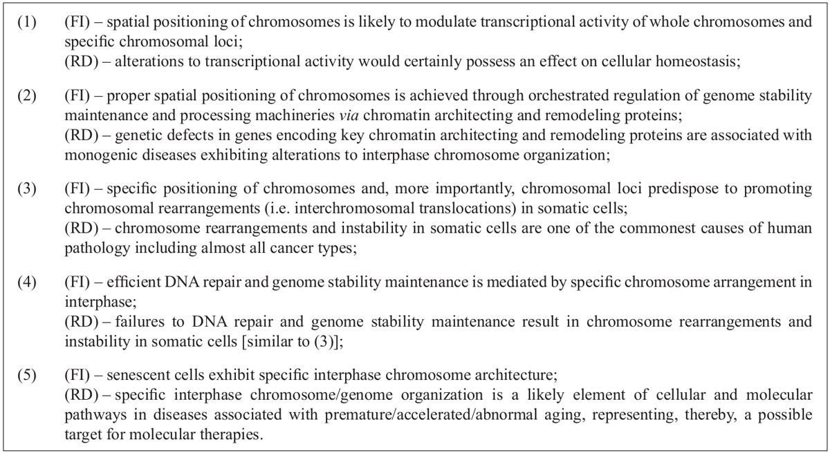
Functional implications (FI) of interphase chromosome organization and its possible relevance to disease (RD).

Interphase chromosome architecture plays an important role in modulation of transcriptional activity through chromatin organization [15] and regulation of cellular and developmental pathways [16]. Therefore, it is not surprising that a number of diseases associated with genetic defects in genes encoding chromatin architecting and remodeling proteins [17] as well as diseases characterized by genome and/or chromosome instability (i.e. cancers) are hallmarked by alterations to spatial genome organization in interphase nuclei [18]. These are suggested to result from failure of genome maintenance and DNA repair machineries, which also depend on interphase chromosome architecture [19]. On the other hand, specific chromosome positioning (intermingling of chromosome territories) is considered as a mechanism of promoting cancer-causing interchromosomal translocations [8, 10, 11, 18]. Furthermore, interphase chromosome associations (somatic pairing) seem to be involved in regulation of transcriptional activity within specific chromosomal regions including imprinted genomic loci, known to be linked to hereditary diseases and cancer [20]. Another critical process that is featured by specific interphase chromosome behavior is the programmed cell death [21]. Since this phenomenon is a key step in numerous pathogenic processes, a link between alterations to interphase chromosome architecture and pathological programmed cell death appear to exist. Finally, the expanding complexity of genomic landscape in senescent cells implies the change of nuclear genome (chromosome) organization as a mechanism for aging at cellular level [22]. The latter has been partially confirmed by direct evaluations [23].

Although several positive associations between specific interphase chromosome architecture and critical nuclear processes have been made, it is usually hard to come to a definite conclusion concerning pathogenic value of variable chromosome arrangement in interphase nuclei. More probably, specific interphase chromosome organization is rather an element of a pathogenetic pathway rather than a unique underlying disease cause. This idea is further supported by observations on diseases caused by mutations in genes encoding chromatin architecting and remodeling proteins [17] and generation of cancer-causing chromosomal rearrangements [18]. Therefore, the analysis of nuclear genome organization defines disease pathways being more complex than previously recognized and an association of abnormal cellular phenotype (or disease phenotype) with specific nuclear genome organization seems to be an important milestone in understanding molecular mechanisms of human pathology.

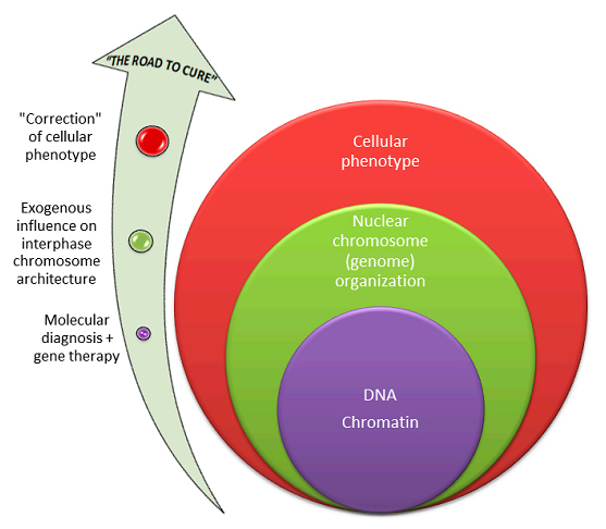
Spatiotemporal interphase chromosome organization is highly dynamic [14] and is able to be exogenously manipulated [24, 25]. Considering that exogenous influences changing interphase chromosome architecture are far from being completely determined [25], one can already speculate on possible applications of manipulating nuclear structure. Specific interphase chromosome organization can be thus defined as a dynamic element of a pathogenetic pathway, which can be influenced. Such changes would certainly have an impact on genome transcriptional activity and stability maintenance, resulting in a beneficial effect and underlying a hypothetic molecular mechanism for “chromosome-oriented” therapy. Hypothesizing a part of such roadmap to “cure” diseases, in which a positive association with specific interphase chromosome/genome organization is made, is given in Figure 1. Nonetheless, it is to be stressed that these assumptions require further studies by means of high-resolution molecular cytogenetic interphase techniques and it is hard to disagree with Dr. Liehr and his colleagues [10] that further studies are necessary for delineation of interphase architecture inalmost all diseases demonstrating abnormal chromosome behavior.

Figure 1
Figure 1. Hypothesizing a part of a suggested roadmap to “cure” diseases associated with specific nuclear organization: (i) according to current concepts in biomedicine, genomic/epigenomic changes occurring at DNA or chromatin levels (purple), which can result in abnormal interphase chromosome architecture, require molecular diagnosis and might be “managed” by gene therapy; (ii) nuclear chromosome (genome) organization (green) can be manipulated exogenously being, therefore, a target for therapies against alterations to interphase chromosome architecture, which are likely to be elements of disease pathways; (iii) at the cellular level (red), the phenotype of a cell would be corrected, if (i) and (ii) were performed successfully.

Concluding remarks

Dr. Liehr and colleagues [10] have drawn the attention of BioDiscovery’s readers to the importance of studying interphase chromosome architecture and its associations with pathological conditions. Taking into account the interest to high-order genome organization, positive data seem to be required for encouraging further attempts at characterization of nuclear genome organization in health and disease. Moreover, technological performance of the study, which defines the value of such associations, demonstrates ICS-MCB as the promising method of choice for studying interphase chromosomes at molecular resolution. The anticipated success of related studies is highly dependent on the way how spatial arrangement of interphase chromosomes is determined. Future studies implementing ICS-MCB for evaluation of nuclear genome have the potential to shed light on the role that interphase chromosome architecture does play in disease.

Acknowledgments

I would like to express my gratitude to Professors Svetlana G. Vorsanova and Yuri B. Yurov for their valuable contributions to and helpful discussions during the preparation of this commentary. The author is supported by RFBR grant 12-04-00215-а (Russian Federation, 2012-2014).

References

  • Misteli T: Beyond the sequence: cellular organization of genome function. Cell 2007; 128: 787-800.
    Reference link
  • Rajapakse I, Groudine M: On emerging nuclear order. J Cell Biol 2011; 192: 711-721.
    Reference link
  • Vorsanova SG, Yurov YB, Iourov IY: Human interphase chromosomes: a review of available molecular cytogenetic technologies. Mol Cytogenet 2010; 3: 1.
    Reference link
  • Iourov IY, Liehr T, Vorsanova SG, Kolotii AD, Yurov YB: Visualization of interphase chromosomes in postmitotic cells of the human brain by multicolour banding (MCB). Chromosome Res 2006; 14: 223-229.
    Reference link
  • Iourov IY, Liehr T, Vorsanova SG, Yurov YB: Interphase chromosome-specific multicolor banding (ICS-MCB): a new tool for analysis of interphase chromosomes in their integrity. Biomol Eng 2007; 24: 415-417.
    Reference link
  • Manvelyan M, Hunstig F, Mrasek K, Bhatt S, Pellestor F, Weise A et al.: Position of chromosomes 18, 19, 21 and 22 in 3D-preserved interphase nuclei of human and gorilla and white hand gibbon. Mol Cytogenet 2008; 1: 9.
    Reference link
  • Manvelyan M, Hunstig F, Bhatt S, Mrasek K, Pellestor F, Weise A, et al.: Chromosome distribution in human sperm - a 3D multicolor banding-study. Mol Cytogenet 2008; 1: 25.
    Reference link
  • Manvelyan M, Kempf P, Weise A, Mrasek K, Heller A, LierA et al.: Preferred co-localization of chromosome 8 and 21 in myeloid bone marrow cells detected by three dimensional molecularcytogenetics. Int J Mol Med 2009; 24: 335-341.
  • Klein E, Manvelyan M, Simonyan I, Hamid AB, Guilherme RS, Liehr T et al.: Centromeric association of small supernumerary marker chromosomes with their sister-chromosomes detected by three dimensional molecular cytogenetics. Mol Cytogenet 2012; 5: 15.
    Reference link
  • Othman M, Lier A, Junker S, Kempf P, Dorka F, Gebhart E et al.: Does positioning of chromosomes 8 and 21 in interphase drive t(8;21) in acute myelogenous leukemia? BioDiscovery 2012; 4: 2.
    Reference link
  • Meaburn KJ, Misteli T, Soutoglou E: Spatial genome organization in the formation of chromosomal translocations. Semin Cancer Biol 2007; 17: 80-90.
    Reference link
  • Iourov IY, Vorsanova SG, Yurov YB: Chromosomal variation in mammalian neuronal cells: known facts and attractive hypotheses. Int Rev Cytol 2006;249: 143-191.
    Reference link
  • Rouquette J, Cremer C, Cremer T, Fakan S: Functional nuclear architecture studied by microscopy: present and future. Int Rev Cell Mol Biol 2010; 282: 1-90.
    Reference link
  • Strickfaden H, Zunhammer A, van Koningsbruggen S, Köhler D, Cremer T. 4D chromatin dynamics in cycling cells: Theodor Boveri’s hypotheses revisited. Nucleus 2010; 1: 284-297.
    Reference link
  • Schneider R, Grosschedl R: Dynamics and interplay of nuclear architecture, genome organization, and gene expression. Genes Dev 2007; 21: 3027-3043.
    Reference link
  • Ktistaki E, Garefalaki A, Williams A, Andrews SR, Bell DM, Foster KE et al.: CD8 locus nuclear dynamics during thymocyte development. J Immunol 2010; 184: 5686-5695.
    Reference link
  • Misteli T: Higher-order genome organization in human disease. Cold Spring Harb Perspect Biol 2010; 2: a000794.
    Reference link
  • Lever E, Sheer D: The role of nuclear organization in cancer. J Pathol 2010; 220: 114-125.
  • Misteli T, Soutoglou E: The emerging role of nuclear architecture in DNA repair and genome maintenance. Nat Rev Mol Cell Biol 2009; 10: 243-254.
    Reference link
  • Göndör A, Ohlsson R: Chromosome crosstalk in three dimensions. Nature 2009; 461: 212-217.
    Reference link
  • Bártová E, Jirsová P, Fojtová M, Soucek K, Kozubek S: Chromosomal territory segmentation in apoptotic cells. Cell Mol Life Sci 2003; 60: 979-990.
  • Yurov YB, Vorsanova SG, Iourov IY: GIN’n’CIN hypothesis of brain aging: deciphering the role of somatic genetic instabilities and neural aneuploidy during ontogeny. Mol Cytogenet 2009; 2: 23.
    Reference link
  • Mehta IS, Figgitt M, Clements CS, Kill IR, Bridger JM: Alterations to nuclear architecture and genome behavior in senescent cells. Ann N Y Acad Sci 2007; 1100: 250-263.
    Reference link
  • Foster HA, Bridger JM: The genome and the nucleus: a marriage made by evolution. Genome organisation and nuclear architecture. Chromosoma 2005; 114: 212-229.
    Reference link
  • Mateos-Langerak J, Goetze S, Leonhardt H, Cremer T, van Driel R, Lanctôt C: Nuclear architecture: Is it important for genome function and can we prove it? J Cell Biochem 2007; 102: 1067-1075.
    Reference link